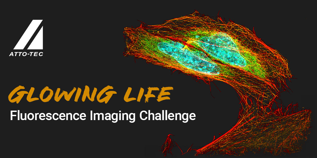
Discover the beauty. Capture the detail. Share your expertise. Win the prize.
We are seeking the most striking microscopic images showcasing ATTO-TEC dyes in action. Whether you are using our latest dye kits or any other ATTO-TEC dye, your task is to create a photo that highlights the beauty and complexity of scientific exploration at its smallest scales.
By clicking submit, you agree to the Terms and Conditions.
Closing Date 13.08.2024
 Cellular Rollercoaster Danush Taban, University of Wuerzburg
This image depicts an 8-fold expanded COS7 cell that has been transfected with 3xGFP-EMTB, a construct where the microtubule-binding domain of ensconsin (EMTB) is fused to three copies of green fluorescent protein (GFP). The GFP is labeled with an anti-GFP nanobody conjugated to ATTO 643. Panel A) displays the GFP signal, Panel B) shows the ATTO 643 labeling, and Panel C) the overlay of both.
Category: Any ATTO Dye
 Julia Weingart, University of Wuerzburg, Department of Biophysics
Cosmic cell nebula - Actin filaments stained with Phalloidin-ATTO565 (white) and whole proteom staining with an ATTO643-NHS ester (magenta) of COS7 cells. The image was taken on a Zeiss LSM900 AiryScan2 setup at 63x magnification. Scale bar: 10µm
Category: Any ATTO Dye
Link on Linkedin Beyond imaging limits Stefan Sachs, University of Wuerzburg - Department of Biotechnology
ATTO-TEC: Glowing life Fluorescence imaging challenge U2OS cells imaged with Lattice-SIM (Zeiss Elyra 7) and 63xoil objective. Whole proteome is shown in grey (ATTO 647N), MitoTracker in orange and alpha-tubulin in green (ATTO 488). DNA of the nuclei is stained with Hoechst (blue). Merged super-resolution image is shown on the right. Scale bars: 10 microns
Category: Any ATTO Dye
Link on Linkedin The Hidden Life of Colon Karen Dubois Camacho, University Medical Center Groningen
Expansion microscopy was used to expand a paraffin-embedded mouse colon tissue section. The gel was stained for whole proteome with Atto643-NHS (green) and with Hoechst for nuclei staining (magenta). Image acquisition: Zeiss LSM900 Airyscan2 microscope, 40x Lens. Enterocytes (left), a goblet cell (right), infiltrating cells (below). Brightest green signal shows vesicles and cell-cell interactions.
Category: Any ATTO Dye
Link on Linkedin HeLa eating nucleotides Cedric Cappel, Institute of Biochemistry, Kiel University
This image shows the uptake of a small oligonucleotide, double-tagged with ATTO 590 and ATTO 655 (purple) by HeLa cells into lysosomes, confirmed by immunostaining against the lysosomal protein LAMP2 with AlexaFluor 488 (green), precisely depicting the limiting lysosomal membrane. A counterstain with DAPI highlights the nuclear DNA (blue). The image was taken at a Zeiss LSM 980 Airyscan 2.
Category: Any ATTO Dye
Link on Linkedin ATTO-TEC meets Pop Art Marcel Streit, Rudolf-Virchow Center, University Würzburg
The image shows COS cells transfected with the human angiotensin-converting enzyme (ACE) receptor. An unnatural amino acid was incorporated into the ACE receptor via Genetic Code Expansion, enabling site-specific labeling. The inserted unnatural amino acid was subsequently clicked with a terazine dye, Me-Tet ATTO-655, allowing for fluorescent visualization of the ACE receptor on the membrane. Acti
Category: Any ATTO Dye
Link on Linkedin Stefan Sachs, University of Wuerzburg - Department of Biotechnology
Immunofluorescence of DIV14 hippocampal mouse neurones and astrocytes. Vesicular glutamate transporter 1 (VGLUT1) is stained with ATTO643 labeling kit. Actin filament staining (orange) is visualized with Phalloidin-ATTO565. Neurofilament L is shown in green with ATTO488 labeling kit. DNA staining (cyan) via Hoechst 34580. Images were taken at 40X magnification on a Zeiss LSM900 Airyscan2 setup.
Category: ATTO Labeling Kits
Link on Linkedin Spirals in the Brain Janna Eilts, Biocenter, University of Wuerzburg
Spiraling Through the Brain: Taenidial Ridges in the Drosophila Brain This Structured Illumination Microscopy image presents a slice of a Drosophila brain after undergoing an 8-fold Expansion Microscopy approach and labeling with NHS-Atto643. While this staining method targets the entire proteome, in this case it especially highlights the taenidia (part of the tracheal system) very efficiently.
Category: Any ATTO Dye
Link on Linkedin Count the organelles Ignacio Vega Vasquez, Biozentrum, University of Wuerzburg
The membrane probe mCLING conjugated with Atto647N was used in mice hippocampal cultures as an approach to visualize endoplasmic reticulum cisterns in the soma of the neurons. The image is from a culture that was subjected to an expansion microscopy protocol, achieving ~8.3 physical expansion of the specimen, before acquiring the image in an LSM 900 Airyscan 2 Zeiss microscope.
Category: Any ATTO Dye
Link on Linkedin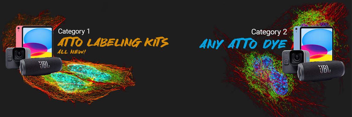
Take a micrograph using at least one ATTO-TEC dye. There are two categories you can enter:
Category “ATTO Labeling Kits” for those using our new Antibody Labeling Kit.(Buy kits with a 20% discount here)
Category “Any ATTO Dye” for images with any other ATTO-TEC dye. Prizes are awarded for both categories individually.
Post your photo with the hashtags #attotec and #GlowingLifeChallenge on LinkedIn to earn extra points through our “Audience Choice Bonus”. Encourage your network to like your post. The more likes you gather, the more points you add to your tally, boosting your chances in the contest.
Complete the contest submission form with your details. Remember, we may verify the use of ATTO-TEC dyes, so be ready to provide proof. To activate the Audience Bonus, don't forget to include the direct link to your LinkedIn post.
After submitting, check your inbox for a confirmation email from us. Click the confirmation link within 48 hours to finalize your contest entry.
1st Place: 10th Generation iPad in the color of your choice
2nd Place: GoPro Hero 11 mini
3rd Place: JBL Flip 6 Outdoor Soundbox
By clicking submit, you agree to the Terms and Conditions.
Closing Date 13.08.2024